Muscles and Nerves of the Orbit
AUDIENCE
Medical students
MEDIA
Adobe Photoshop
Description
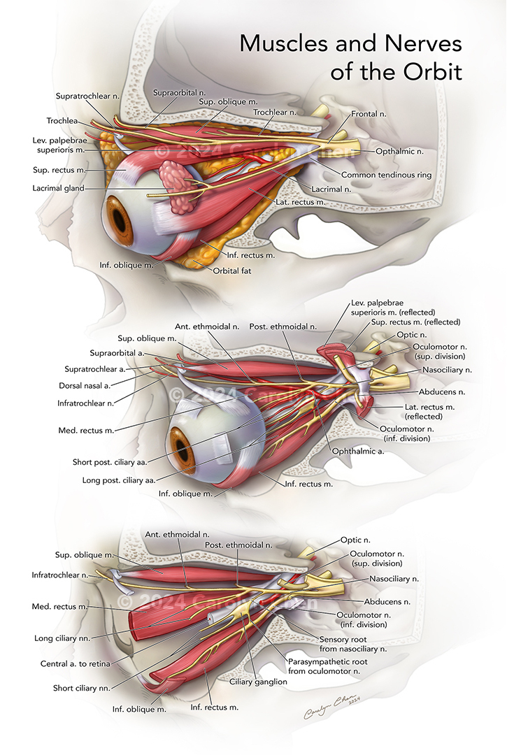
This anatomical plate illustration depicts the normal anatomy of the orbit as a sequence of images revealing the muscles, nerves and arteries from superficial to deep. The top image establishes a superior oblique view with a section of the skull removed to reveal the left eye and orbit. In the middle image, the lateral rectus, superior rectus, and levator palpebrae superioris muscles have been reflected to reveal the optic nerve and the nerves superficial to it. The ophthalmic artery and its branches are included to show its superior-lateral course around the optic nerve. In the bottom image, the eye has been removed to reveal the medial and inferior rectus muscles and the attachment between the inferior oblique muscle and orbital cavity. The lateral half of the common tendinous ring was also removed to show the relationships between the nerves as they enter the orbit. To ensure contrast and legibility, all text is above a 4.5:1 luminance ratio, and the color palette accommodates color blindness.